Pregnancy ultrasound scans: how many and what they show
The question I get asked most about scans isn’t “what will they see” — it’s “how many is too many?” Somebody has had a third scan booked in six weeks and is quietly panicking that all that sound energy is doing something. Somebody else has had exactly two and is convinced they’re being short-changed.
Both worries are reasonable, and both have a boring answer: it depends on where you live, why the scan was booked, and whether it’s answering a real clinical question. So let’s do the safety question first, because it’s the one that keeps people awake, and then walk through the schedule scan by scan. ❤️
Are multiple ultrasounds safe?
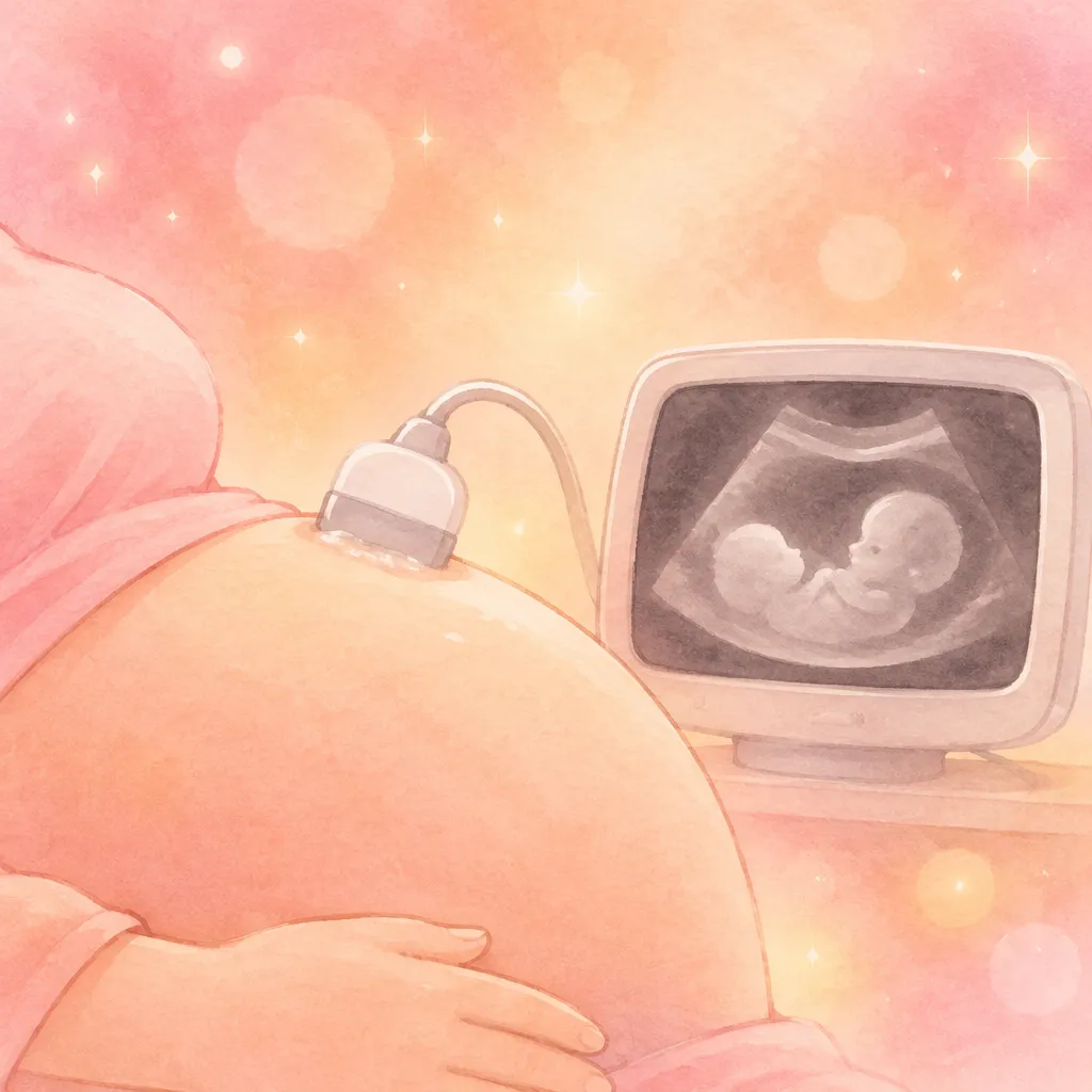
Ultrasound is not an X-ray. It uses high-frequency sound waves, not ionising radiation, and it’s been used in pregnancy for more than forty years without a demonstrated risk at diagnostic levels. That’s the honest, evidence-based position, and it’s the one ACOG, the NHS and the WHO all take.
Two things worth understanding, though:
- It isn’t “zero energy.” The beam slightly warms tissue and produces tiny mechanical vibrations. That’s why guidelines follow the ALARA principle — as low as reasonably achievable. A trained sonographer doing a diagnostic scan stays well inside safe limits. A 40-minute 4D “keepsake” session in a shopping centre is a different animal.
- There’s no universal maximum number. You’ll see “no more than three” quoted online, usually without a source. Guidance doesn’t work that way. A twin pregnancy with growth restriction might get a scan every two weeks, and that’s appropriate care, not over-testing.
ACOG specifically advises against non-medical, keepsake-only ultrasound — the kind where you pay for a DVD of your baby’s face. Not because a single session is dangerous, but because there’s no clinical benefit to weigh against prolonged exposure.
So: if your midwife or doctor books another scan, ask what it’s for. Nine times out of ten there’s a clear reason. If there genuinely isn’t one, you’re allowed to ask why.
How many scans you’ll actually get (it depends on the country)
This is where most of the confusion comes from, because “normal” changes at the border.
| Where | Routine scans | Weeks |
|---|---|---|
| UK (NHS) | 2 | Dating scan 11-14, anomaly scan 18-21 |
| US | 1-2 typical | Dating/viability 7-12 if unsure of dates, anatomy scan 18-22 |
| Australia (Medicare) | 2-3 | Dating 8-13, nuchal/first trimester 11-13+6, morphology 18-22 |
| Germany, Italy, Spain | 3 | One per trimester |
| Anywhere, if indicated | As needed | Growth scans, Doppler, twins, diabetes, hypertension |
Two scans is genuinely the NHS standard, and it surprises a lot of people who expected one per trimester. It isn’t rationing — it’s that the two scans with the strongest evidence behind them are the dating scan and the anomaly scan. Everything else is added when there’s a reason.
Private scans and “peace of mind” bookings
Private scan clinics have exploded, and I understand why: a 10-week scan after a previous loss, or a reassurance scan before telling family, can be worth every penny emotionally. Just keep two things in mind. First, a private scan is not a substitute for your NHS or insurance-covered anomaly scan — it doesn’t replace the detailed 20-week check. Second, the person scanning should be a qualified sonographer who will report findings properly, not just print a photo and wave you out.
The dating scan: why it matters more than people expect
Booked between roughly 11 and 14 weeks, this one does three jobs at once.
- It dates the pregnancy. Crown-rump length at this stage estimates your due date to within about five days — far more accurate than counting from your last period, especially if your cycles are irregular.
- It counts the babies and the placentas. Twins change your entire care pathway, and whether they share a placenta changes it again.
- It’s the window for the first-trimester screening test (the combined test: nuchal translucency plus a blood test). That window closes at 14 weeks and 1 day, which is why this scan is booked when it is.
A note on the nuchal translucency measurement you’ll see on the report: it’s a screening marker, not a diagnosis. A thicker measurement means a higher chance, not a condition. If the combined screening comes back higher-chance, the next step is a discussion about NIPT or a diagnostic test — not a conclusion.
The 20-week scan: what the anatomy scan is really checking
If you only remember one scan, remember this one. The anomaly scan — usually between 18 and 21 weeks — is a systematic top-to-toe check, and it takes 30-45 minutes when the baby cooperates.
What’s on the checklist:
- Brain and spine: ventricles, cerebellum, spinal cord, skin covering the spine.
- Face: lips, palate, eye sockets, profile.
- Heart: four chambers, outflow tracts, heart rate and rhythm.
- Abdominal wall, stomach, kidneys, bladder: confirms organs are where they should be.
- Arms, legs, hands, feet: long bones measured, digits counted.
- Placenta, cord and amniotic fluid: position, cord vessels, fluid volume.
- Cervical length, in some services, as a preterm birth marker.
”They found a marker” — what that actually means
This is the moment that ruins a good day, so let’s be precise. A soft marker is a finding that’s more common in certain chromosomal conditions but also appears in plenty of healthy babies. The usual suspects: an echogenic focus in the heart, choroid plexus cysts, mild kidney dilation (pyelectasis), a slightly short femur, echogenic bowel.
An isolated soft marker is not a diagnosis. It’s a data point that gets weighed alongside your screening results. Most people who get referred for a follow-up scan after a soft marker end up with a healthy baby — you just never hear those stories, because nobody posts “the marker meant nothing” in a forum at 2am.
Do you have to find out the sex?
No, and you can say so at the start of the appointment. In the UK, sonographers generally won’t tell you before 16 weeks anyway, and some NHS trusts have policies about when and whether they’ll confirm it. If you don’t want to know, say it as you walk in — it’s a two-second sentence that saves an awkward moment.
Growth scans in the third trimester
After 26-28 weeks, scans change purpose: nobody is looking for structural problems any more, they’re checking how the baby is growing and how they’re lying.
- Estimated fetal weight and centile. Calculated from head, abdomen and femur measurements — and accurate to roughly plus or minus 10-15%. That margin is why a single “big baby” or “small baby” reading doesn’t decide anything.
- Presentation. Head down, breech or transverse. Plenty of babies turn after 32 weeks; the conversation about options comes later if they don’t.
- Placenta position. If it was low at 20 weeks, this is when they check whether it has moved up.
- Amniotic fluid volume. Too little or too much changes monitoring.
- Doppler, if growth is a concern: it measures blood flow resistance in the umbilical artery and tells you how well the placenta is working.
The key idea: growth is a trend, not a snapshot. One low centile with a normal curve is usually nothing. Two scans showing the curve flattening is what gets acted on.
What the scan names mean
| Term | What it is | What it’s for |
|---|---|---|
| Dating scan | Standard first-trimester scan | Confirm viability, count babies, set the due date |
| NT / nuchal scan | Measurement of the fluid at the back of the neck | Part of first-trimester screening |
| Anomaly / anatomy scan | Detailed structural scan at 18-22 weeks | Checks organs, growth, placenta |
| Growth scan | Third-trimester measurement scan | Tracks weight centile, fluid, position |
| Doppler | Blood-flow study of umbilical/uterine arteries | Placental function in higher-risk pregnancies |
| 3D / 4D | Volume rendering, still or moving | Keepsake images; limited diagnostic value |
| Transvaginal | Internal probe | Most reliable before about 10-11 weeks |
“4D” and “5D” are marketing labels on top of the same physics. They do not detect anything a 2D scan misses — the diagnostic work happens in 2D, always.
How to prepare, and what to ask
- Early abdominal scans usually want a full bladder — drink water about 30 minutes before.
- Transvaginal scans want the opposite: empty bladder.
- Wear two pieces, so you can lift a top rather than undress.
- Bring your notes and your questions. Write them down; they evaporate in the room.
- Ask about photos and recording when you book — policies vary wildly.
- Ask who reports the result and when. In the NHS it goes to your midwife or consultant; privately, make sure you know who reads it.
Good questions to have ready: Is the growth measuring on track for my dates? Is the placenta where it should be? Do you need to see me again, and why? Who will contact me if something needs following up?
When not to wait for the next appointment
Scans are scheduled, but symptoms aren’t. Contact your midwife, maternity triage or emergency services the same day if you have:
- Heavy bleeding (soaking a pad) or bleeding with clots, with or without pain.
- Severe one-sided abdominal pain, especially with shoulder-tip pain or feeling faint — that combination needs urgent assessment.
- Any fluid leaking from the vagina, a gush or a constant trickle.
- Reduced or absent fetal movements after 24 weeks, for more than 12 hours.
- Severe headache, visual disturbance or sudden swelling of hands and face — possible pre-eclampsia.
- Fever with feeling unwell, or vomiting so severe you can’t keep fluids down.
If you’re playing the waiting game before your first scan and want something to do with your hands, our online pregnancy test quiz is a symptoms-based starting point (orientational only), and the finger pregnancy test game is exactly what it sounds like — a game, not a test. For dates, the week-by-week hub and how many weeks pregnant am I will tell you more than a scan photo ever will about where you are right now.
Frequently asked questions
How many ultrasounds do you get in a normal pregnancy?
In the UK, two on the NHS: the dating scan at 11-14 weeks and the anomaly scan at 18-21 weeks. In the US it’s commonly one to two, depending on whether a dating scan was needed. In Germany, Italy and Spain it’s usually one per trimester, so three. Extra scans are added when there’s a clinical reason.
Can too many ultrasounds harm the baby?
No evidence of harm has been found at diagnostic exposure levels, and ultrasound doesn’t use ionising radiation. There’s no fixed maximum number — what matters is whether each scan has a medical purpose. Long, non-medical keepsake sessions are the ones guidelines advise against, because there’s no benefit to balance against the exposure.
Can you tell the sex at the 12-week scan?
Not reliably. The “nub theory” angle people analyse on forums is genuinely unreliable at that stage, which is why most sonographers won’t guess. The 20-week scan is the usual point for that, and even then it depends on the baby’s position. NIPT blood testing can determine sex much earlier, but it’s offered for screening purposes, not as a gender-reveal service.
What happens if they find something at the anatomy scan?
You’ll be told what was seen and what it might mean, and referred onward if needed — often to fetal medicine for a more detailed scan, sometimes for a diagnostic test. Many findings turn out to be isolated markers with no condition behind them. Try not to search the finding the same evening; context-free information is frightening by design.
Why does the estimated weight change so much between scans?
Because it’s an estimate built from indirect measurements, with a real margin of error of roughly 10-15%. The baby’s position changes what can be measured, and different sonographers measure slightly differently. The trend across scans is what clinicians act on.
Do I need a full bladder for the scan?
For an early abdominal scan, yes — a full bladder lifts the uterus into a better position, so you’ll usually be asked to drink water beforehand. For a transvaginal scan, you’ll be asked to empty it. If nobody tells you, ask when you book.
Is the 20-week scan ever wrong?
It can miss things — no scan detects everything, and some conditions only become visible later in pregnancy. It’s a screening test, not a guarantee. That’s also why the anomaly scan is done by a trained sonographer with a structured checklist rather than a quick look.
Do I have to drink water and hold it for the whole appointment?
Usually just for the scan itself. If your bladder is too full and uncomfortable, say so — the sonographer can often work around it, or let you partially empty it.
Sources
- ACOG — Ultrasound exams (FAQ)
- NHS — Ultrasound scans in pregnancy
- NHS — 12-week scan
- NHS — 20-week scan
- MedlinePlus — Fetal ultrasound
- WHO — Antenatal care (fact sheet)
- NICE — Antenatal care (NG201)
This article is for information only and does not replace your doctor’s or midwife’s assessment. If you have any concerns about your pregnancy, pain, bleeding or reduced movements, always speak to a healthcare professional ❤️